Day 6: In full bloom
By Michelle Cusolito
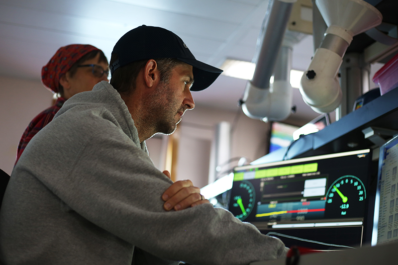
It’s obvious we’ve traveled north because the sun rises earlier now than it did in Spain. By 7am, the main lab is already filled with bright light that reflects off the waves outside. Co-chief scientist Heidi Sosik has been awake all night—but you would never know it. She smiles as she stares at her computer screen.
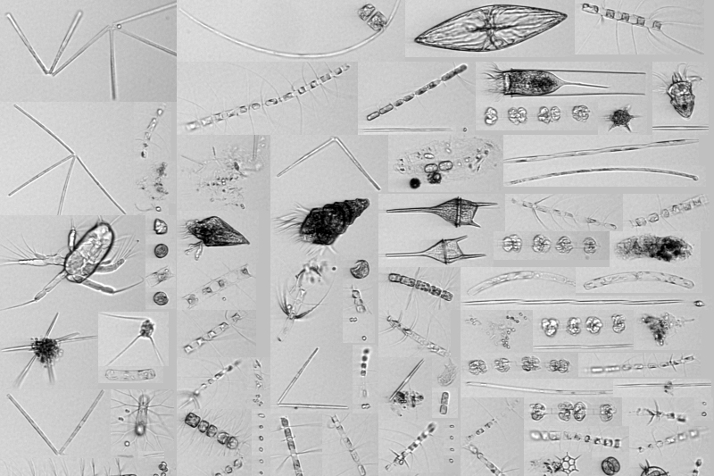
“This is unlike anything I’ve ever seen,” she exclaims. “Sarmiento de Gamboa is in an absolute diatom soup!”
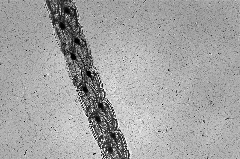
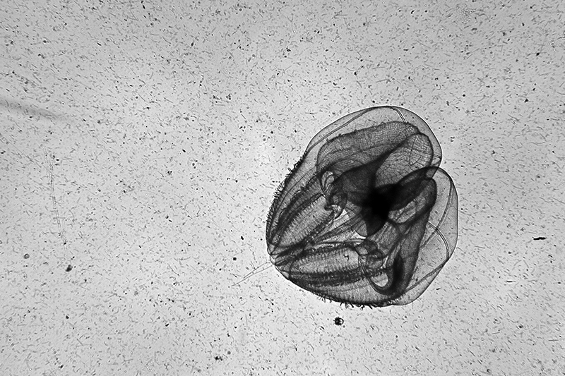
She is referring to the images of diatoms flashing in front of her. These images are being collected by the shadowgraph camera system, known as the In-situ Ichthyoplankton Imaging System (ISIIS). Towed behind the ship on a “sled” called StingRay, a fiberoptic cable sends images and other data back to scientists on board.
ISIIS is designed to image larger organisms than diatoms, a type of phytoplankton that measures (at most) about the width of a human hair. But Heidi is confident she sees diatoms because she compared the images from the shadowgraph to the high-resolution images coming from the Imaging FlowCytobot (IFCB). Heidi and a colleague invented the IFCB to measure the details of diatoms and other phytoplankton. IFCB pumps seawater through a tube and a laser triggers the camera to take a photo of each particle right when it is in focus.
The abundance of diatoms suggests an active bloom may be underway—exactly what the science team was hoping to find in this area of the North Atlantic.
“When the spring bloom is ending and phytoplankton die, we expect to see lots of carbon falling from the sunlit zone toward the deep ocean,” Heidi explains. “It’s like when tree leaves fall. They land on the ground and decay. When phytoplankton die, they sink too.”
As phytoplankton sinks deeper into the ocean, some are eaten by hungry zooplankton. When that grazing animal poops or dies, the carbon in its last meal sinks further into the ocean. As the carbon works its way through the food chain, some of it will end up on the ocean floor, where it may be trapped in sediments for centuries. Scientists onboard the Sarmiento de Gamboa are trying to figure out exactly what happens to carbon in the deep sea, to better understand the ocean’s role in regulating Earth’s climate.
While the images collected by the IFCB and ISIIS provide important scientific information, they also serve as entertainment for all of us who enjoy looking at ocean life. Not all data sets are visually compelling, but the images generated by these sophisticated instruments have an artistic quality to them, almost like finely-detailed charcoal drawings.
“When you shine light on gelatinous animals, their delicate edges and inside parts cast beautiful shadows,” Heidi explains. “We take pictures of those shadows to help identify these fragile creatures and where they live in the ocean.”
National Geographic photographer David Liittschwager, who joins the expedition on assignment with the magazine, is also impressed.
“I stayed up too late last night watching the images come in from Heidi’s instrument—it’s mesmerizing,” he says. “There’s this incredible quantity of life. Images go by at 14 frames per second, and if you look close enough, there is something alive in nearly every frame, even down deep.”
David isn’t alone. Many of us stayed up too late last night and will likely do the same many nights over the coming weeks. How can we sleep when ground-breaking science is happening in front of our eyes?


