|
December 6, 2011 Slideshow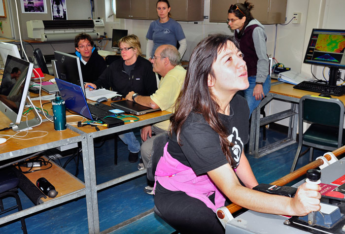
Whenever the SID-ISMS went to the depths, scientists gathered in the computer lab to send it commands, read the data coming back from its sensors, and monitor its performance. Here, Maria Pachiadaki controls the winch that is lowering the instrument toward the Deep Hypersaline Anoxic Basin. Behind her, scientists Thorsten Stoeck, Ginny Edgcomb, and Craig Taylor watch the data that SID-ISMS is sending to the ship. Behind them, SSSG Catie Graver and scientist Hera Karayanni observe. 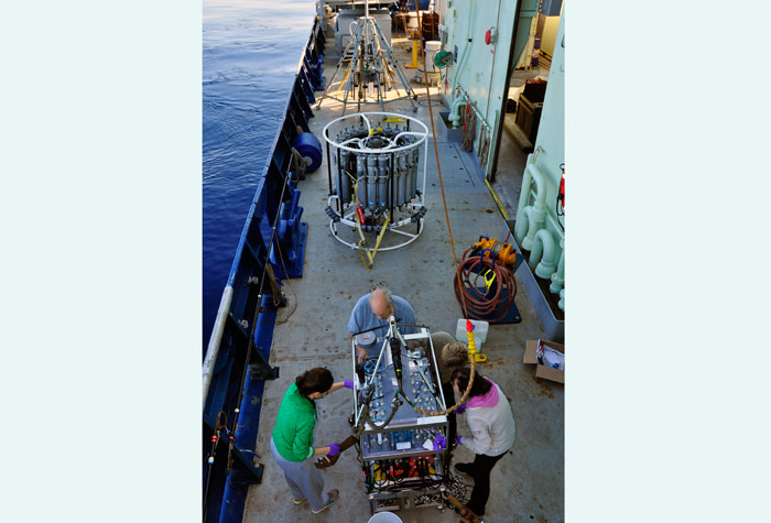
(clockwise from left) Hera Karayanni, Craig Taylor, Ginny Edgcomb, and Maria Pachiadaki gathered around SID-ISMS after every deployment to gather samples and assess what went right and what went wrong. Behind them are the CTD rosette and (way in the back) the MC800 multicorer. 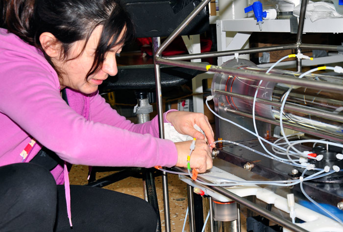
Maria Pachiadaki starts assembling Deep SID. All the tubing had to be connected to the correct ports and flushed with distilled and deionized water. 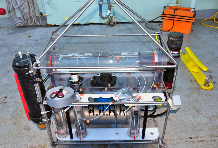
Deep SID is less than half the size of SID-ISMS and cannot go as deep or perform as many tasks, but it can fix (preserve) water samples while up to 1,000 meters below the surface of the ocean. It works by taking a 4-liter sample of water into the horizontal chamber on the top, then pumping portions of the large sample into the individual sample tubes arranged vertically below. 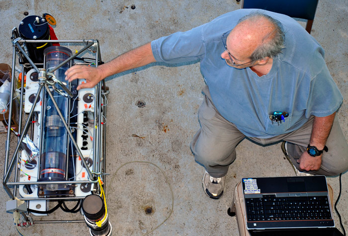
Craig Taylor gets reacquainted with Deep SID as he prepares to program it for the sampling tasks ahead. He is the co-designer of both Deep SID and SID-ISMS. Deep SID must be programmed before the dive. SID-ISMS can respond to orders sent to it during the dive by scientists on the ship. 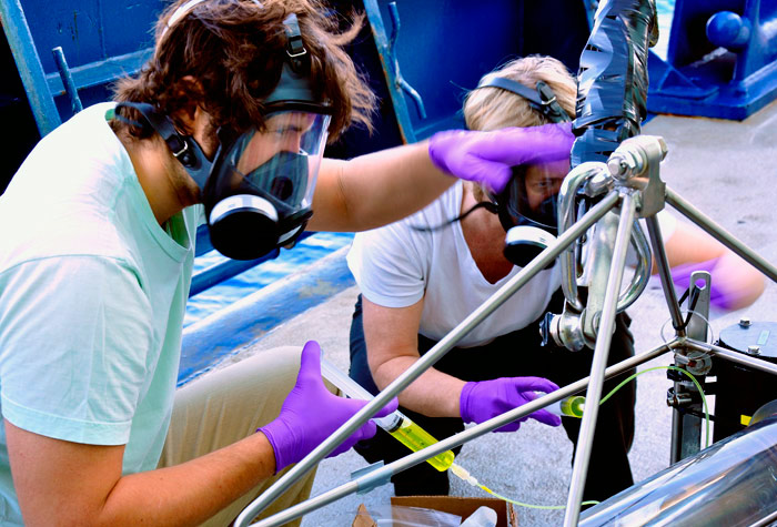
Bill Orsi and Ginny Edgcomb begin to load Deep SID’s sample chambers with bright yellow Bouin’s fixative, which will preserve microbes in the water for microscopic examination. They are wearing masks to protect them from fumes from the fixative. 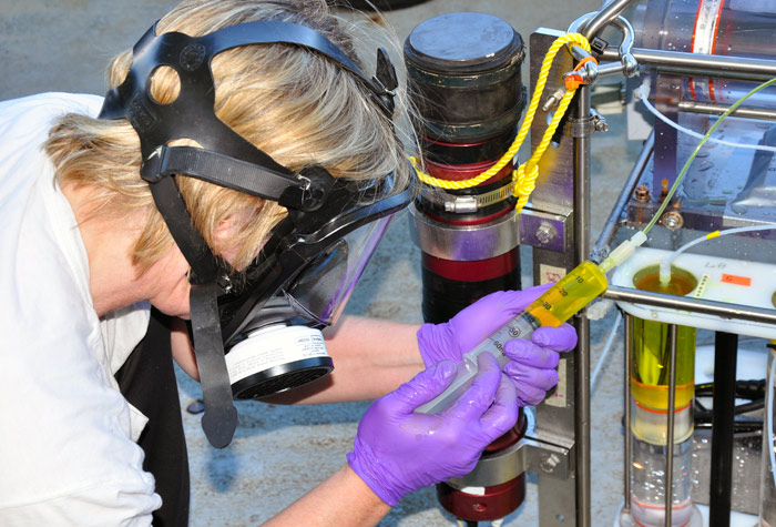
Ginny Edgcomb injects Bouin’s fixative into one of Deep SID’s sample chambers. At the start of a deployment, the top half of each chamber contains fixative and the bottom half contains water. A piston separates the two halves. During a deployment, sample water flows into the top of the chamber, mixing with the fixative, forcing the piston down, and pushing the pre-loaded water out of the chamber 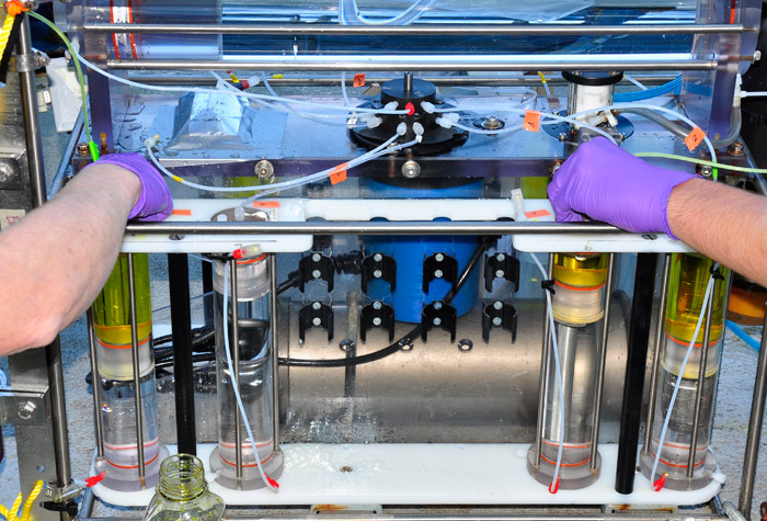
When Ginny Edgcomb and Bill Orsi finish loading the Deep SID, all four chambers will have yellow Bouin’s fluid at the top and water at the bottom. 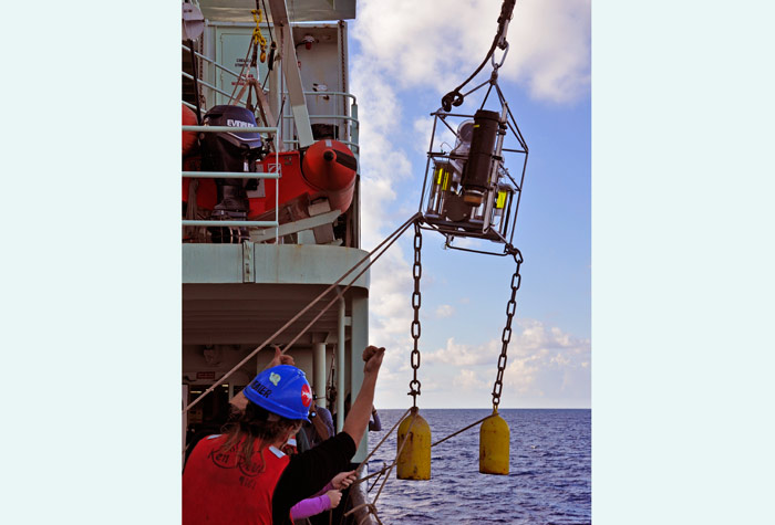
Deep SID heads out on a test deployment. The heavy “bullets” hanging from its frame help the lightweight instrument remain vertical in the water. 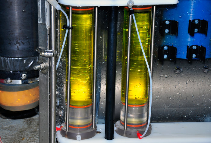
Success! During a deployment at 40 meters depth, Deep SID performed perfectly. One sign of that is that the yellow Bouin’s fixative, which started out in the top half of each chamber, is now evenly distributed throughout each chamber. 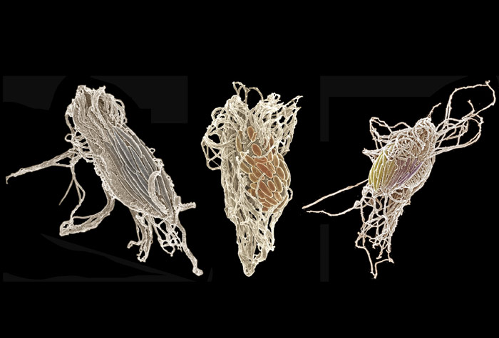
This is the kind of specimen and image that in situ fixation of samples can provide—three protists covered with bacterial symbionts. In each image, the long, thin filaments are flagella of the protist. The more oval shapes are the bacteria. Color was added to the images to make the bacteria easier to see. Bill Orsi and Ginny Edgcomb collected these protists from the Cariaco Basin in Venezuela using Deep SID and viewed them through a scanning electron microscope. Photomontage by Bill Orsi.
|

Mailing List | Feedback | Glossary | For Teachers | About Us | Contact
© 2010 Dive and Discover™. Dive and Discover™ is a registered trademark of Woods
Hole Oceanographic Institution
